MRM Insights: Antler “skin” provides a velvety path towards scarless wound healing
Dr. Irah King
Every month, in the MRM Insights, a member of the MRM Network writes about stem cells and regenerative medicine from a different perspective. This month, Dr. Irah King, Associate Professor in the Department of Microbiology and Immunology at the RI-MUHC, Director of the McGill Centre for Microbiome Research and Canada Research Chair in Barrier Immunity, discusses how antler “skin” provides a velvety path towards scarless wound healing.
Antler “skin” provides a velvety path towards scarless wound healing
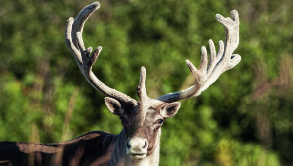
When a recent paper about the regenerative power of Reindeer antler skin was published in Cell, it seemed like a perfect topic for an MRM Insight contribution. After all, growing up in rural Pennsylvania, I was told (and believed) that these wingless creatures possessed the power of flight and could silently land on my rooftop (for the record, I no longer endorse that idea). While they may not be magical, reindeer are majestic, in part because of their incredible ability to grow massive, spike-like bones out from the top of their head (see the photo I took from my office window below). Called antlers, these towering hat racks can rise up to 4 feet in length, making these seemingly gentle creatures quite fierce. Although antlers serve as weapons for territorial and mating dominance, these multi-pronged pokers literally fall off and grow back every year (talk about a regenerative network!). As part of this annual rebirth, antlers are covered with velvet, a highly vascularized, innervated skin-like layer containing hair follicles and all. For the aficionados, the term velvet derives from the latin velveeta (pronounced vel-vee-tuh) which was said to be a delicious, silky cheese-like product made by the ancient Mesopotamians. Seriously, look it up! Many of us will also be familiar with the term in reference to the texture of your grandmother’s musty couch that you weren’t allowed to sit on as a child. OK, enough reminiscing, what can antler velvet teach us about regeneration??!!
Since antlers are rapidly growing, semi-calcified bones that are exposed to the outside world, velvet provides a protective coat that is shed when the animals reach mating age and the antlers are fully calcified. Based on the remarkable ability of reindeer to re-grow their antlers and velvety cover each year, a research group at the University of Calgary led by Dr. Jeff Biernaskie, a world leader in tissue regeneration and fibroblast biology, posited that velvet may possess unique regenerative properties compared to other mammalian skin. Moreover, they hypothesized that velvet may even scarlessly heal wounds (Sinha et al, Cell, 2022)1. If they were correct, this would be a huge discovery because most wounds in mammals leave a scar (there are exceptions – cue the spiny mouse2) and this would be the first description of different regenerative pathways in the same tissue (i.e. skin) from the same animal! Importantly, it could also pave the way for identifying mechanisms of scarless healing in a model very similar to human skin.
To test their hypothesis, the authors induced full-thickness wounds in the velvet and back skin of adult reindeer living within an outdoor enclosure at the University of Calgary Veterinary Research Station. When they quantified how the wounds healed, they found that while the back skin formed a scar, the healing antler velvet became indistinguishable from uninjured velvet! To quantify the regeneration, the authors measured hair follicle density at the wound site (scars don’t regrow hair follicles). Indeed, the velvet wound bed recovered 73% hair follicle density compared to only 12% in the back skin wound. Remarkably, they obtained similar results when they performed full-thickness burn injuries on a different group of reindeer. These results provided a powerful setting to reveal the mechanisms underlying the different regenerative properties of velvet versus back skin. But how could these different mechanisms be revealed? Well, the authors took a page out of the old transplant research playbook and grafted velvet onto the back skin of the same reindeer and repeated the incisional wound in the velvet patch. For comparison, they also injured native (on antler) velvet from the same animal. Remarkably, the transplanted velvet also healed with no scar! These findings led the authors to do a deep dive into velvet that would even make Lou Reed proud. Specifically, they wanted to know what cells in the velvet (i.e. velvet cells: very cool) were responsible for scarless healing. For this, they performed single-cell RNA sequencing (scRNAseq) on total cells isolated from the velvet and back skin. Maybe not too surprisingly, they found that gene expression in fibroblasts was the most different between the velvet and back skin. Given that fibroblasts are central to tissue remodeling and wound repair3 they examined this population more carefully. Interestingly, velvet fibroblasts expressed a “regenerative” transcriptional signature, under steady-state conditions, while back skin fibroblasts expressed genes related to inflammation. Of note, the most highly expressed genes in velvet cells related to regeneration (e.g. CRABP1 and MDK) are part of the Vitamin A/retinoic acid signaling cascade, a pathway known to support both hematopoietic4 and mesenchyme-derived stem cell5 populations. In addition, they found an association of scarring (fibrotic) healing with Yap target gene expression, a mechano-sensitive transcriptional activator of the Hippo pathway critical for tissue regeneration (as our very own Yap expert, Alex Gregorieff, can tell you6).
Following these observations, Sinha et al. performed an elegant set of kinetic studies using scRNAseq and scATACseq experiments on skin over two weeks post-wounding and demonstrated similarities between the initial tissue response at the different sites but that back skin fibroblasts adopted a later form of inflammatory, myofibroblast-driven contractile healing (skin contraction leads to scarring) that never occurred in the antler velvet. Rather, the velvet retained much of their baseline regenerative gene signature throughout the scarless healing process. Given the inflammatory nature of the back skin scarring response, the authors also examined differences in immune cell infiltration and coupled this with in vitro primary fibroblast cultures to understand cell crosstalk following wounding. These kinetic and co-culture studies revealed another fundamental insight into wound healing: fibroblasts from different skin sources modulate the function of incoming immune cells that, in turn, shape the modes by which fibroblasts mediate tissue repair.
As if these advances weren’t enough, Sinha et al. wanted to more generally understand why the regenerative capacity of fetal skin is lost with age. To tackle this question, they performed an extended kinetic analysis of their velvet skin transplants. By measuring hair follicle density in these grafts over a six-month period, they discovered that while “young” wounds regained follicles at a density similar to non-grafted velvet, hair follicles significantly diminished as the wounds “aged”. Employing machine learning approaches on scRNAseq data to gain insight into these temporal changes, Sinha et al. identified a population of fetal-like fibroblasts that expressed a “transitionary” phenotype between regenerative and inflammatory states. Over time, these transitionary fibroblasts responded to tissue stress signals (likely via Yap again) and started to produce immunostimulatory factors including the chemokine CXCL12 and macrophage growth factor CSF1 that, via communication with immune cells, promoted fibroblasts to adopt a fibrotic phenotype. Obviously, the most important conclusion from these studies is that it’s all about the immune system*.
*Disclaimer: I’m an immunologist
Overall, I found this study not only fun to read but also incredibly informative regarding the fundamental mechanisms of wound healing. As such, I drew two main conclusions from this landmark paper: 1. Fibroblasts are seminal players in the outcome of wound healing, not only by establishing the baseline regenerative capacity of a tissue, but also by shaping the impact of the immune cells that come calling in response to injury and 2. Fibroblasts are much less boring than I thought. Moreover, this research represents bona fide “outside of the box” thinking. After all, the research group had no experience using reindeer as a model system, but they were brave enough to pursue their question anyway. I’m sure the story behind the idea is as interesting as the science itself. As any good research project does, this discovery provokes many more questions. For example, what are the developmental pathways that determine the baseline epigenetic signature of velvet versus back skin fibroblasts? What are the critical immune populations that shape the fibroblast response following injury? Can we harness this knowledge (pun intended) to improve the treatment of patients with severe burns? In any case, my childlike fascination with reindeer has been restored!
References
1. Sinha, S. et al. Fibroblast inflammatory priming determines regenerative versus fibrotic skin repair in reindeer. Cell 185, 4717-4736 e4725 (2022).
2. Seifert, A.W. et al. Skin shedding and tissue regeneration in African spiny mice (Acomys). Nature 489, 561-565 (2012).
3. Plikus, M.V. et al. Fibroblasts: Origins, definitions, and functions in health and disease. Cell 184, 3852-3872 (2021).
4. Cabezas-Wallscheid, N. et al. Vitamin A-Retinoic Acid Signaling Regulates Hematopoietic Stem Cell Dormancy. Cell 169, 807-823 e819 (2017).
5. Batourina, E. et al. Vitamin A controls epithelial/mesenchymal interactions through Ret expression. Nat Genet 27, 74-78 (2001).
6. Gregorieff, A. & Wrana, J.L. Multiple roles for the hippo effector yap in gut regeneration and cancer initiation. Mol Cell Oncol 3, e1143992 (2016).